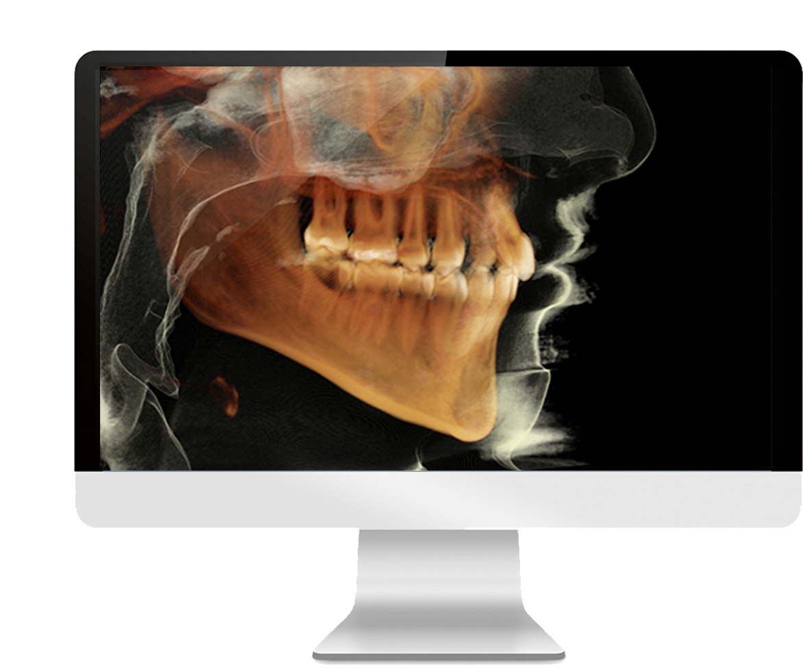
Cone Beam CT: Cone beam computed tomography is one of the most significant advancements in oral and maxillofacial imaging. Using imaging equipment that rotates around the head, this technology works with advanced computerized technology to generate 3-D images of teeth and the maxillofacial area (mouth, jaw and neck). These 3-D views provide dentists with information on the patient's anatomy, as well as the extent of a disease or condition, thereby helping in diagnosis and treatment planning.
Applications of CBCT imaging include, but are not limited to:
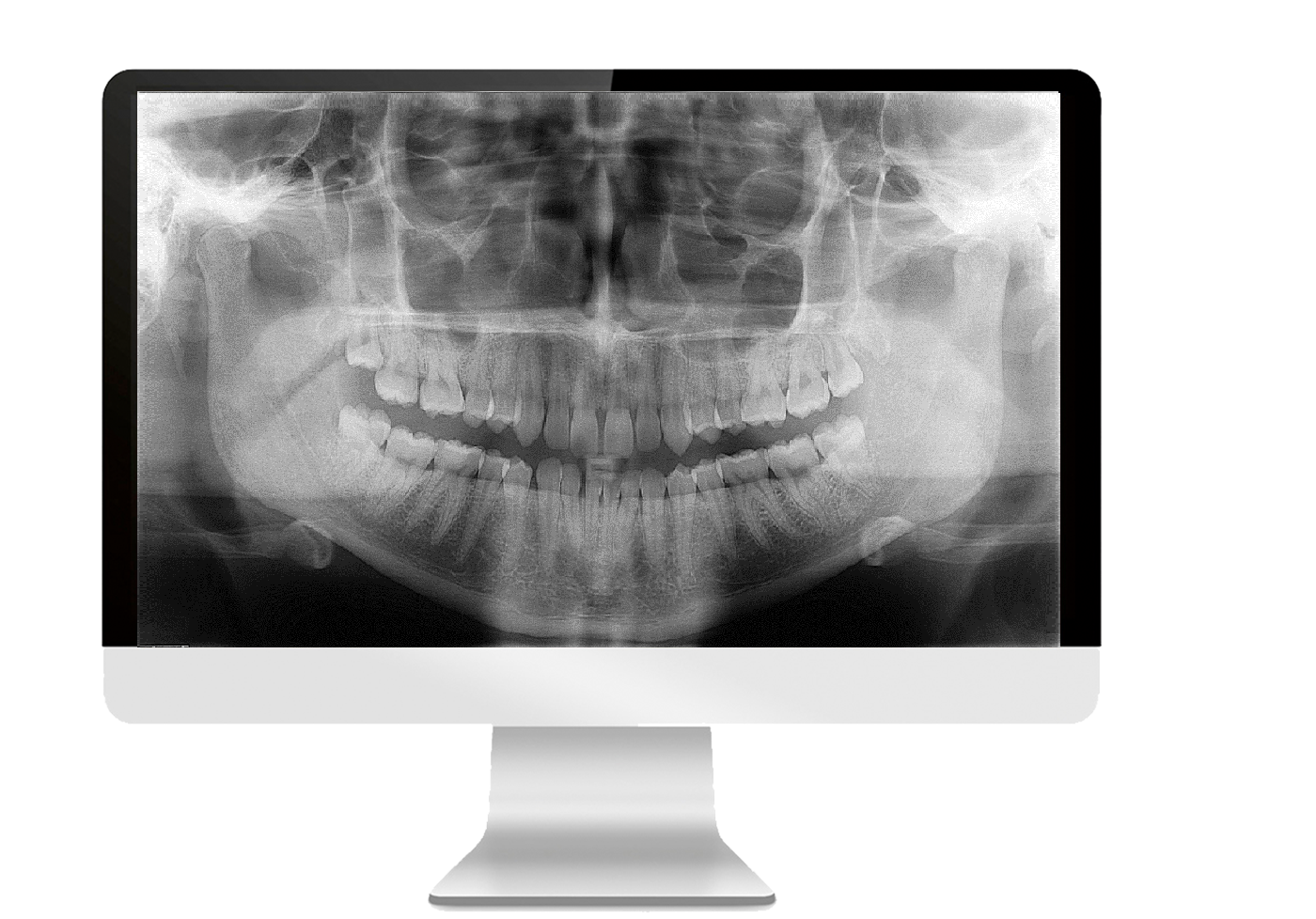
Radiographic exams commonly used to image teeth, jaws, cranium, and cervical spine by placing an image receptor (film or sensors) outside the patient's mouth.
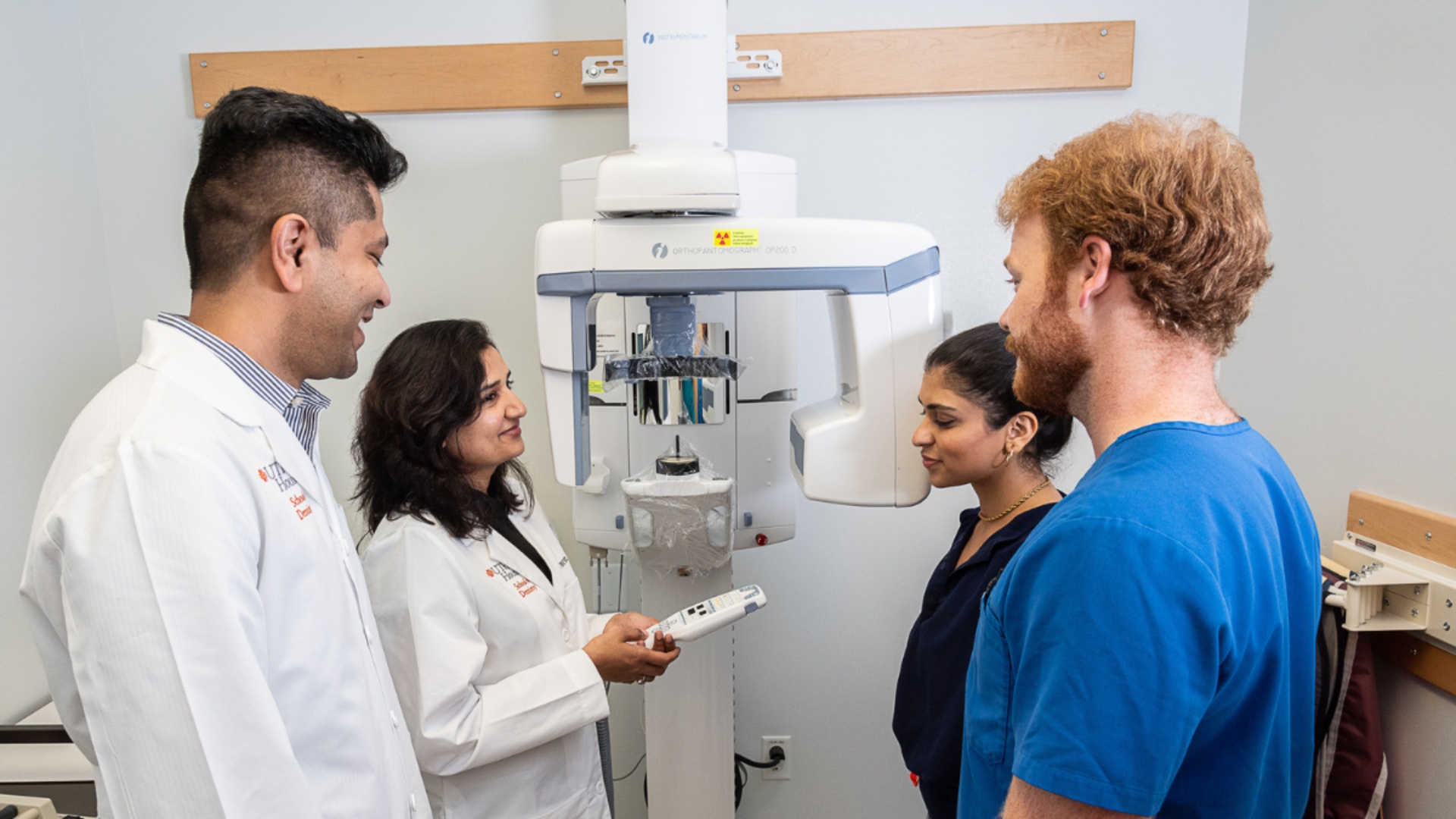
We perform the following sets of imaging:
A faculty oral and maxillofacial radiologist at UT Dentists Imaging will interpret the images and prepare a report that addresses the primary purpose of the case and includes a detailed pathology assessment, along with key images. Radiologists will be available to answer referring doctors’ questions and concerns regarding the case.
Reports are completed within three business days; rush service is available on a case-by-case basis. UT Dentists Imaging uses a HIPAA-compliant image-sharing portal to make reports and images readily accessible any time at your convenience.
We also provide specialized services, such as airway analysis, skeleton-maturation analysis and TMJ MRI interpretation.
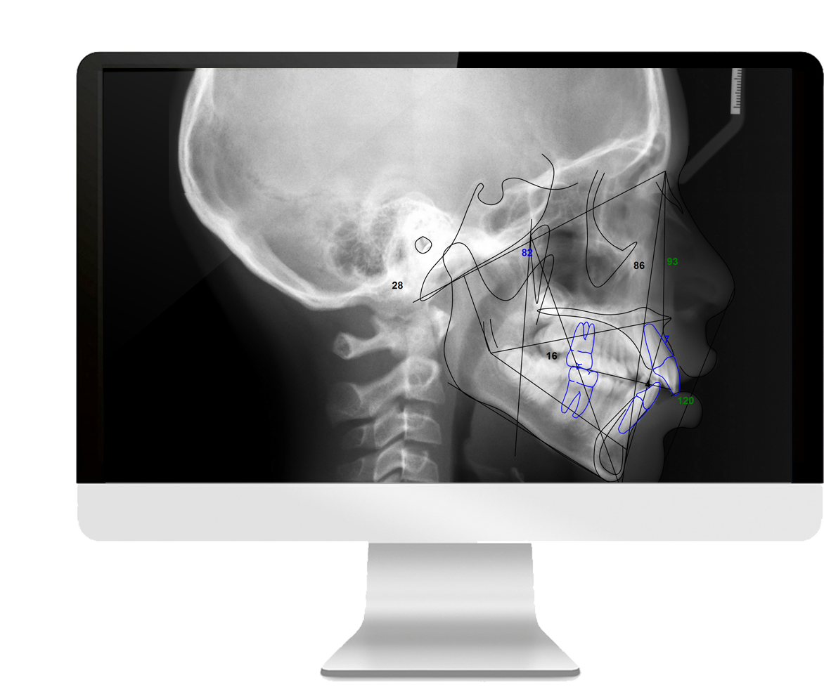
2-D imaging often poses diagnostic challenges due to superimposition of anatomical structures, variations in projection geometry or — on certain occasions — due to positioning and technical errors. Our oral and maxillofacial radiologists are here to help and overcome diagnostic challenges by offering interpretation services for 2-D extraoral radiographs, and to make further recommendations when needed.
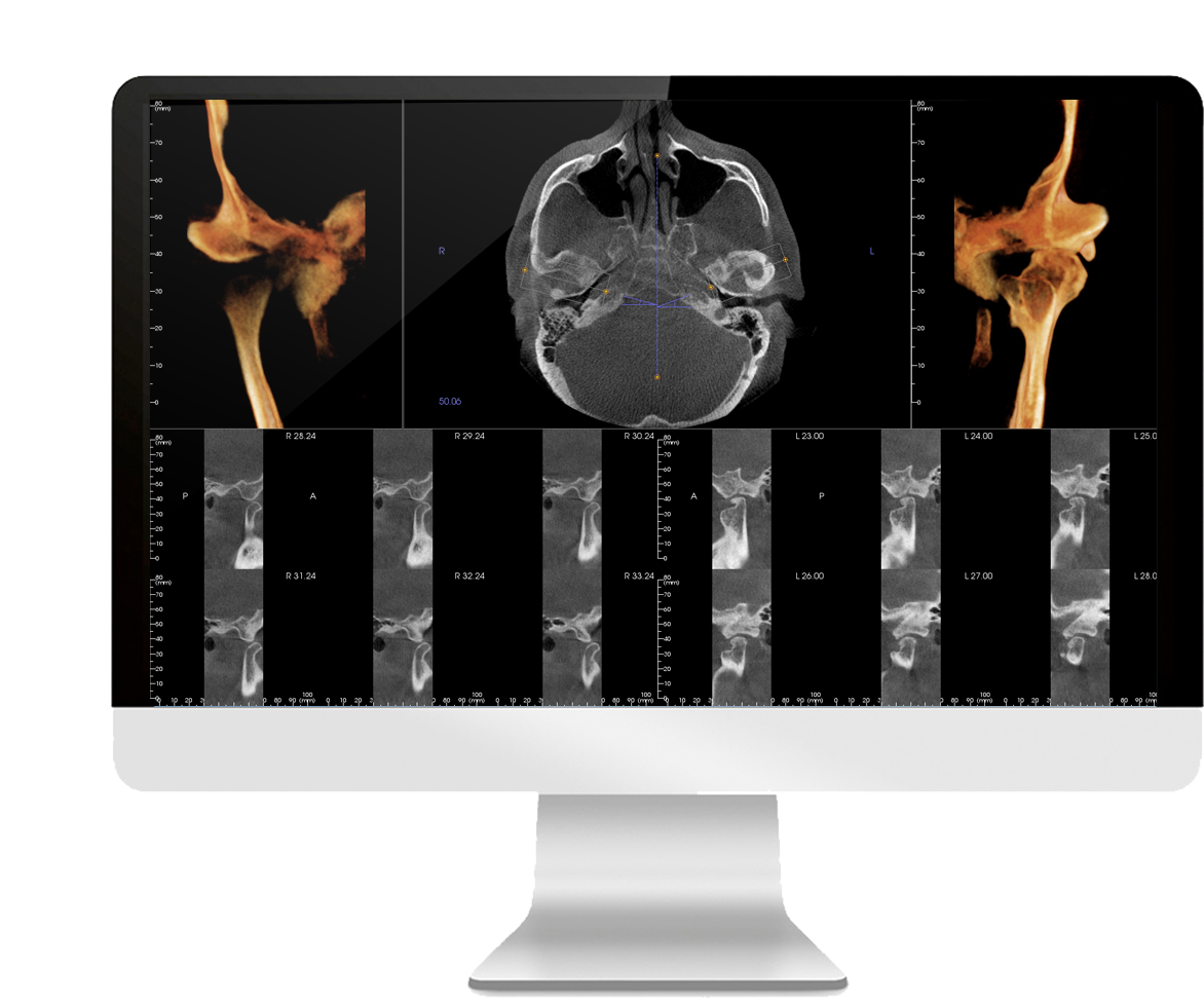
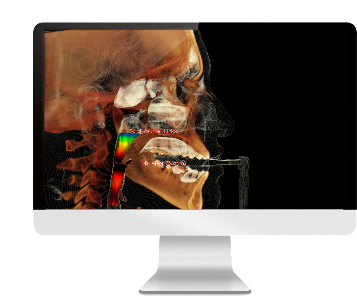
Cone Beam CT can be used effectively to assess the upper airway in patients with dentofacial discrepancies, malocclusion and obstructive sleep apnea. Our oral and maxillofacial radiologists perform complex assessments of CBCT data on a variety of software types to segment and perform volumetric measurement of the upper airway.
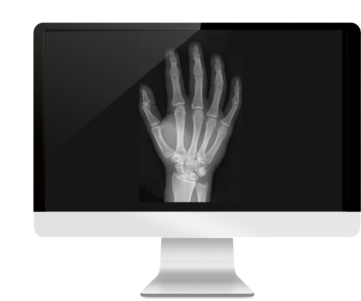
Skeletal maturation patterns vary with individuals and often pose challenges for clinicians performing orthodontic interventions. Hand-wrist radiography is considered one of the oldest methods for maturity indicators. Our technologists are experienced in performing digital hand-wrist radiography, and our oral and maxillofacial radiologists can provide a detailed assessment of skeletal maturation.